
Specialist care, nerve microsurgery and the patient pathway at CHU de Nice (Pasteur 2 Hospital).
1. What is the brachial plexus?
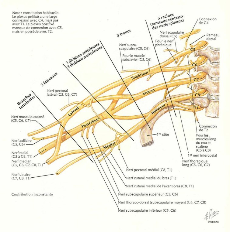
The brachial plexus is a complex network of nerves located at the base of the neck and at the shoulder. It can be compared to a set of « electrical cables » connecting the spinal cord (itself connected to the brain, which is the « electrical generator ») to the various muscles and areas of skin of the upper limb.
These nerves control all movement and sensation in the arm:
- The shoulder: to stabilise the joint and raise the arm.
- The elbow: to bend and straighten the arm, allowing the hand to be brought to the face.
- The hand and wrist: to provide grip (closing the fingers) and protective sensation.


2. Causes and mechanisms of brachial plexus injuries
Traumatic brachial plexus palsies occur most often in young adults (18-30 years), most frequently following a high-energy motorcycle or road traffic accident. The mechanism of injury is a violent stretching between the neck and the shoulder.
The two main types of injury
- Nerve rupture (« the broken cable »): the nerve is interrupted along its course and scar tissue (a neuroma) forms, blocking the transmission of nerve impulses.
- Nerve avulsion (« the plug torn out at the source »): the nerve root is torn directly from the spinal cord at the level of the cervical spine.
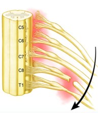
Clinical presentation and symptoms
The palsy may be total (an upper limb that is completely flail and without sensation) or partial (selectively affecting the shoulder, elbow or hand). Neuropathic pain (burning, electric shock-like pain) frequently accompanies the injury.
3. The diagnostic work-up at CHU de Nice
Prompt care within the Institut Universitaire Locomoteur et du Sport (IULS), at Pasteur 2 Hospital, requires a rigorous preoperative work-up:
- Specialist clinical examination: rigorous segment-by-segment assessment of each muscle group and analysis of associated signs.
- Electromyography (EMG): generally performed from the 3rd to 4th week onwards to analyse the electrical activity of the nerves and muscles.
- Advanced radiological imaging (MRI of the plexus or CT myelography): essential for precisely mapping the anatomical condition of the cervical nerve roots.
4. Principles and strategy of surgical management
Time is critical: the therapeutic window
A muscle deprived of its nerve supply deteriorates irreversibly. The neuromuscular junction (the « motor end plate ») disappears permanently after 18 to 24 months.
This is why nerve microsurgery must be planned within the first 6 months following the injury.
The hierarchy of functional priorities
As the available nerve resources are limited, the surgical strategy carried out by the specialist team follows a strict order of priority to restore independence:
| Priority | Function to be restored | Goal for the patient |
|---|---|---|
| No. 1 (absolute) | Elbow flexion | To allow the hand to be brought to the mouth, face and body. |
| No. 2 | Shoulder stability and mobility | To position the arm in space and stabilise the shoulder girdle. |
| No. 3 | Protective sensation | To protect the hand against repeated injuries and burns. |
| No. 4 | Hand movement | To restore grip and the opening/closing of the fingers. |
5. Advanced microsurgical techniques
A. Early nerve reconstructions (less than 6 months)
Nerve grafts
In the case of a nerve rupture where good-quality roots are preserved, the surgeon bridges the gap by grafting segments of a sensory nerve harvested from the leg (sural nerve), re-establishing a conduction bridge.
Nerve transfers (neurotisations)
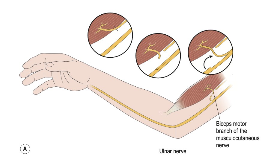
In the case of root avulsion, nerve transfers are used: a nearby functioning donor nerve (or a healthy nerve fascicle) is harvested and connected directly to the paralysed nerve as close as possible to the target muscle, shortening the distance the nerve has to regrow.
- For the elbow: transfer of fascicles of the ulnar nerve or median nerve to the nerve to the biceps muscle, to reanimate elbow flexion.
- For the shoulder: transfer of a motor branch of the triceps to the axillary nerve (deltoid muscle), and transfer of the spinal accessory nerve to the suprascapular nerve to reanimate the rotator cuff.
B. Secondary palliative techniques
When the optimal time window has passed, the team offers palliative procedures:
- Secondary tendon and muscle transfers: rerouting a healthy muscle to take over the lost function.
- Free vascularised muscle transfers (gracilis): harvesting a muscle from the thigh with its vessels and nerve to restore active flexion of the arm.
- Joint fusion (arthrodesis): bone and joint procedures can also help to stabilise the joints and improve certain functions.
For details of each procedure, see the article Brachial plexus surgery.
6. Care pathway and rehabilitation at CHU de Nice
Nerve regrowth takes place at the biological rate of approximately 1 millimetre per day. Postoperative follow-up at CHU de Nice is therefore a long-term process.
The pillars of functional recovery
- Specialist physiotherapy: maintaining joint mobility and preventing stiffness from the very first days.
- Neuromotor re-education: supporting the patient in integrating the new nerve commands at brain level.
- Multidisciplinary care: monitoring of neuropathic pain in conjunction with the specialist units of the CHU.
In summary
- Brachial plexus injuries require highly specialised expertise, which is available within the Institut Universitaire Locomoteur et du Sport (IULS) at CHU de Nice.
- The time to referral is crucial: seek a consultation as quickly as possible if there is any neurological deficit of the upper limb after an injury.
- A dedicated team of surgeons, anaesthetists, pain physicians and rehabilitation specialists supports you throughout your care pathway.
Frequently asked questions about the brachial plexus
What is brachial plexus palsy?
The brachial plexus is a network of nerves located at the base of the neck and at the shoulder, which controls movement and sensation in the arm. After an injury, the palsy may be total (an upper limb that is completely flail and without sensation) or partial (selectively affecting the shoulder, elbow or hand).
What is the most common cause of a brachial plexus injury?
Traumatic brachial plexus palsies occur most often in young adults (18-30 years), following a high-energy motorcycle or road traffic accident. The mechanism is a violent stretching between the neck and the shoulder.
How soon should a brachial plexus injury be operated on?
Nerve surgery must be planned within the first 6 months following the injury. A muscle deprived of its nerve supply deteriorates irreversibly, and the neuromuscular junction disappears permanently after 18 to 24 months.
Which investigations are carried out before the operation?
The work-up combines a specialist clinical examination, electromyography (EMG) generally performed from the 3rd to 4th week onwards, and imaging (MRI of the plexus or CT myelography) to map the condition of the cervical nerve roots.
How long does recovery take after surgery?
Nerve regrowth takes place at a rate of approximately 1 millimetre per day, so follow-up is a long-term process. It combines specialist physiotherapy, neuromotor re-education and management of neuropathic pain.
What can be offered once the 6-month window has passed?
Palliative procedures remain possible: tendon and muscle transfers, free vascularised muscle transfer (gracilis) to restore active flexion of the arm, or joint fusion (arthrodesis).
Where can I be seen in Nice for a brachial plexus injury?
Care is provided within the Institut Universitaire Locomoteur et du Sport (IULS) at CHU de Nice, at Pasteur 2 Hospital. It is advisable to seek a consultation as quickly as possible if there is any neurological deficit of the upper limb after an injury.
Book an appointment: for a consultation with Professor Olivier Camuzard in Nice, go to the Contact page.